Introduction
For over a decade, precision oncology has promised to tailor cancer therapies to the unique molecular vulnerabilities of an individual's tumor. The completion of landmark projects like The Cancer Genome Atlas (TCGA) established a foundational understanding that cancer is fundamentally a disease of the genome [1]. However, as the field matured, it became painfully clear that DNA sequencing alone is insufficient to capture the staggering complexity of tumor biology. A mutation may be present, but whether it drives the cancer, how the immune system reacts to it, and how the tumor physically manifests in the body requires a broader lens.
Historically, oncologists, pathologists, and radiologists have operated in relative isolation, each analyzing a distinct facet of the disease through their own specialized modalities. This siloed approach often leads to fragmented clinical decision-making, where a targeted therapy might be prescribed based on a genetic mutation without considering the spatial context of the tumor microenvironment or the physical boundaries revealed by imaging. The advent of multimodal artificial intelligence (AI) represents a paradigm shift, offering the computational power necessary to synthesize these disparate data streams into a unified, holistic view of a patient's cancer [2].
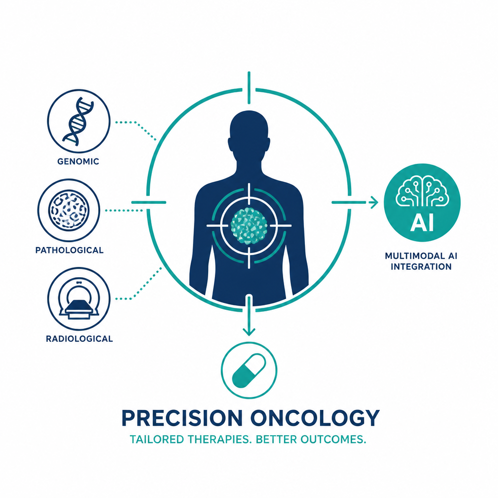
By integrating genomic sequencing, digital pathology, and radiological imaging, multimodal AI models mimic the natural cognitive process of a multidisciplinary tumor board--but with superhuman scale and pattern-recognition capabilities. This article explores how this integration is being achieved, the algorithmic architectures driving it, the transformative clinical applications emerging from it, and the critical hurdles that must be overcome before it becomes standard of care.
The Three Pillars of Oncological Data
To understand the power of multimodal AI, one must first appreciate the unique and complementary nature of the data it consumes. Each modality captures a different "scale" of the tumor.
Genomic Sequencing: The Molecular Blueprint
Genomic data provides the foundational code of the tumor. Next-generation sequencing (NGS) identifies driver mutations, gene fusions, copy number alterations, and mutational signatures. While highly precise, genomics lacks spatial and temporal context. It cannot tell an oncologist how densely packed the cancer cells are, nor can it reveal the physical structure of the surrounding stroma. It represents the "what" of the tumor's potential, but not necessarily the "how" of its current behavior [3].
Digital Pathology: The Microscopic Truth
Whole-slide imaging (WSI) has transitioned pathology from a purely analog to a digital discipline. High-resolution scans of tissue biopsies capture the tumor microenvironment (TME) in exquisite detail--revealing immune cell infiltration, necrosis, and tissue architecture. Deep learning models can extract quantitative morphological features from these slides that are imperceptible to the human eye, known as pathomic features. Pathology bridges the gap between the molecular blueprint and the physical reality of the disease [4].
Radiology and Radiomics: The Macroscopic View
Radiological imaging--such as CT, MRI, and PET scans--provides a non-invasive, three-dimensional view of the entire tumor and its surrounding anatomy. Radiomics, the extraction of mineable, high-dimensional data from medical images, can capture tumor heterogeneity, shape irregularities, and textural patterns. Crucially, imaging allows for longitudinal monitoring, offering dynamic insights into how a tumor evolves in response to therapy over time [5].
The Mechanics of Multimodal Integration
Simply feeding genomic, pathological, and radiological data into a standard neural network does not yield successful multimodal AI. The fundamental challenge lies in "data heterogeneity"--genomics consists of discrete sequences or vectors; pathology is massive, high-resolution image data; and radiology is three-dimensional volumetric data. Developing architectures capable of aligning these distinct representations is the primary technical hurdle in the field [6].
Overcoming the Semantic Gap
Early efforts in multimodal AI relied on "late fusion," where separate unimodal models processed each data type independently, and their final output predictions were averaged or voted upon. While straightforward, this approach fails to capture the complex, cross-modal interactions--for instance, how a specific genetic mutation physically manifests in a radiological texture. Modern approaches favor "intermediate" or "early fusion," where the raw data or high-level feature representations are mapped into a shared, latent multidimensional space [2].
Advanced Algorithmic Architectures
Transformers, the architecture behind large language models, have proven exceptionally adept at this task. By treating genomic sequences, image patches from pathology slides, and spatial voxels from CT scans as distinct "tokens," transformer-based models can learn the complex attention mechanisms that link a KRAS mutation to a specific histological pattern and a corresponding radiomic signature. Furthermore, Graph Neural Networks (GNNs) are being utilized to map the relationships between different biological entities, such as how gene-protein interaction networks influence the cellular architecture visible on a pathology slide [7].
Transformative Clinical Applications
The integration of these modalities via AI is already moving from theoretical research into tangible clinical utilities, fundamentally altering how therapies are selected and evaluated.
Predicting Therapeutic Response with Unprecedented Accuracy
One of the most promising applications is predicting patient response to immunotherapy, which has historically been guided imperfectly by single biomarkers like PD-L1 expression or tumor mutational burden (TMB). Multimodal AI models have demonstrated the ability to combine TMB (genomics), the spatial distribution of CD8+ T-cells (pathology), and tumor heterogeneity (radiology) to predict response to immune checkpoint inhibitors with significantly higher accuracy than any single modality alone [5]. This prevents patients from undergoing toxic, ineffective treatments and allows for rapid pivoting to alternative therapies.
Uncovering Hidden Biomarkers and Subtyping
Multimodal AI excels at unsupervised clustering, identifying entirely new subtypes of cancer that transcend traditional histological classifications. By analyzing the integrated features of thousands of patients, AI can discover that what looks like a standard Grade III glioma under a microscope actually consists of three distinct molecular-morphological subtypes, each with vastly different prognoses and sensitivities to specific chemotherapies [4]. This allows oncologists to reclassify patients based on the true biological nature of their disease rather than arbitrary anatomical classifications.
Navigating the Hurdles: Interpretability and Standardization
Despite its immense potential, the clinical deployment of multimodal AI faces significant non-technical barriers that must be addressed to ensure patient safety and clinician trust.
The Black Box Dilemma and Clinical Trust
The more complex the AI model--particularly deep neural networks and multimodal transformers--the more opaque its decision-making process becomes. In oncology, where life-and-death decisions are made, a "black box" recommendation is insufficient. The field is increasingly focusing on Explainable AI (XAI), developing techniques such as attention mapping and SHAP (SHapley Additive exPlanations) values. These tools allow the AI to highlight the specific region of a pathology slide or the specific gene variant that most heavily influenced its treatment recommendation, translating computational logic into clinical rationale [6].
Standardization and the Promise of Federated Learning
Multimodal AI requires massive datasets to train effectively, but medical data is heavily siloed and subject to stringent privacy regulations like HIPAA and GDPR. Furthermore, variance in imaging equipment, staining protocols, and sequencing platforms creates batch effects that can severely bias AI models. Federated learning is emerging as a critical solution, allowing institutions to train collaborative AI models on their local data without ever sharing the raw patient information. Only the learned model weights are shared, preserving privacy while aggregating the diverse, large-scale data necessary to build robust, generalizable multimodal systems [7].
Conclusion
Precision oncology is undergoing a profound evolution, moving beyond the reductionist view of cancer as a purely genetic disease toward a comprehensive, systems-level understanding. Multimodal AI stands at the vanguard of this transformation, serving as the computational glue that binds together the molecular blueprint of genomics, the cellular reality of digital pathology, and the macroscopic architecture of radiology. While formidable challenges in algorithmic interpretability, data standardization, and clinical validation remain, the trajectory is unmistakable. As these integrative models mature and weave themselves into the fabric of clinical workflows, they will empower oncologists to craft therapies with unprecedented precision--turning the vast, complex data of modern medicine into a highly targeted weapon against cancer.